MAXILLOFACIAL PATHOLOGY
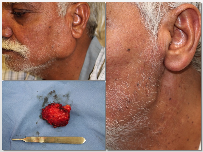

Oral Pathology
Oral pathology covers a broad range of diseases and conditions, which can be either benign (non-cancerous) or malignant (cancerous).
Common conditions –
- Oral Cancer.
- Ulcers
- Benign Lesions
- A description of non-cancerous conditions like fibromas, mucoceles, and hemangiomas.
- Red and white patches in the oral cavity.
- Jaw cysts and tumors
- Salivary gland tumors and infections


Cyst
A cyst is a fluid-filled sac that can develop in the jawbones or soft tissues of the oral cavity.
These cysts are typically slow-growing and may not cause symptoms initially, but they can expand over time, leading to complications.
Common Signs and Symptoms of Oral Cysts:
- Painless swelling in the jaw or gums
- Expansion of the jawbone leading to facial asymmetry
- Displacement or mobility of teeth
- Infection, causing pain and pus discharge in some cases
- Difficulty in chewing, speaking, or swallowing if the cyst enlarges significantly.
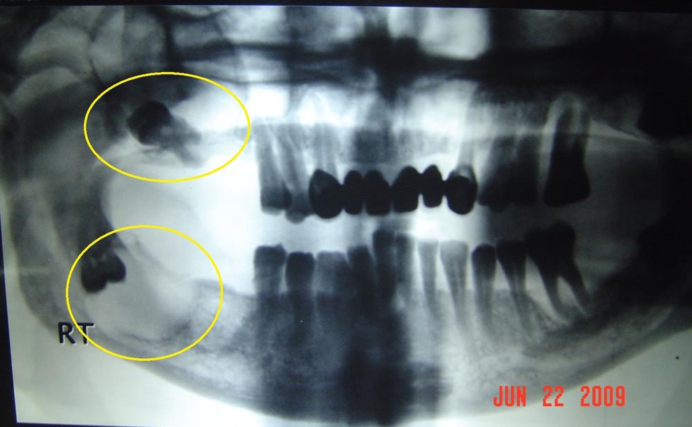
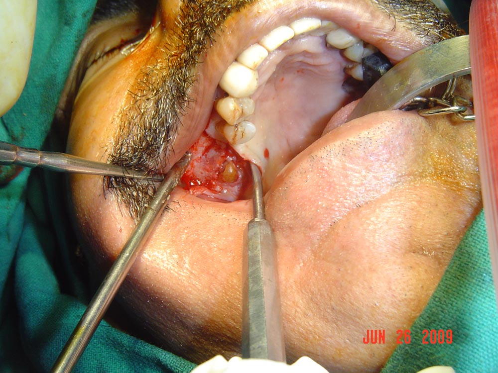

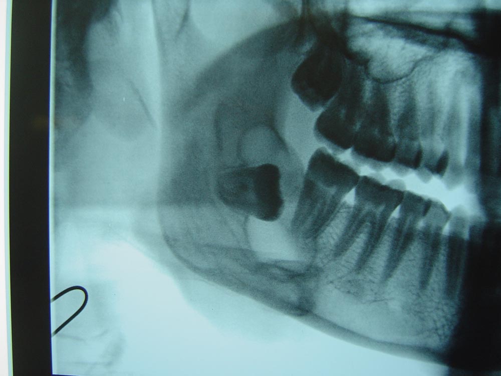


Oral cysts are often detected through radiographic imaging (X-rays), appearing as radiolucent (dark) areas in the jaw. Some cysts remain asymptomatic, while others may cause significant bone destruction or secondary infections if left untreated.
Treatment typically involves surgical removal to prevent further complications such as jaw weakening, fractures, or infections. Regular dental checkups and early diagnosis are essential for effective cyst management and overall oral health.


Tumor
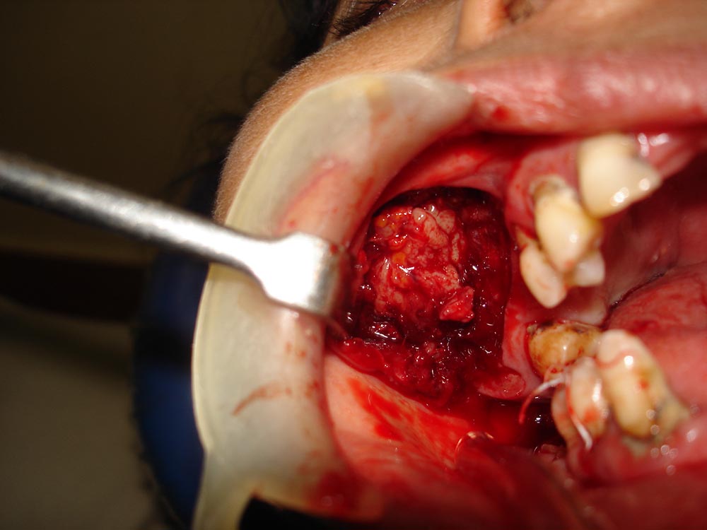
A tumor is an abnormal growth of tissue that can develop in the oral cavity or jaw.
Tumors can be benign (non-cancerous) or malignant (cancerous), with the latter requiring urgent medical attention. Early detection is crucial to prevent complications and ensure effective treatment.
Signs and Symptoms Tumors:
- Persistent swelling or lump in the mouth or jaw
- Unexplained bleeding or ulcer that does not heal
- Sudden mobility of teeth
- Facial asymmetry due to tumor growth
- Pain or difficulty in chewing, swallowing, or speaking
Oral tumors can form in various areas, including the gums, tongue, palate, and jawbones. They are often detected through clinical examination and imaging techniques such as X-rays, CT scans, or MRI. A biopsy is performed to confirm the nature of the tumor and determine the appropriate treatment.
Treatment options include surgical removal, and in the case of malignant tumors, additional therapies such as radiation or chemotherapy may be necessary. Regular dental checkups and early intervention play a vital role in managing oral tumors effectively.
Oral Cancer
Oral cancer is a serious and potentially life-threatening condition characterized by the uncontrolled growth of abnormal cells in the mouth.
It can affect the lips, tongue, cheeks, floor of the mouth, hard and soft palate, sinuses, and throat. Early detection and treatment are crucial for improving survival rates.
Signs and Symptoms of Oral Cancer:
- Reddish (erythroplasia) or whitish (leukoplakia) patches in the mouth
- A sore or ulcer that does not heal and bleeds easily
- Lump, thickening, or rough area inside the mouth
- Difficulty in chewing, swallowing, or speaking
- Persistent sore throat or hoarseness
- Unexplained numbness, pain, or tenderness in the mouth or lips
- Sudden mobility of teeth without an obvious cause.

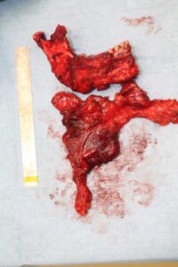

Risk factors for oral cancer include tobacco use (smoking or chewing), excessive alcohol consumption, prolonged sun exposure (lip cancer), HPV infection, and poor oral hygiene.
Diagnosis involves clinical examination, biopsy, and imaging techniques like X-rays, CT scans, or MRIs. Treatment options depend on the stage and severity of the cancer, including surgery, radiation therapy, chemotherapy, or targeted drug therapy.
Regular dental checkups and self-examinations are key to early detection. If any suspicious signs persist for more than two weeks, immediate medical consultation is recommended.



Soft tissue cyst
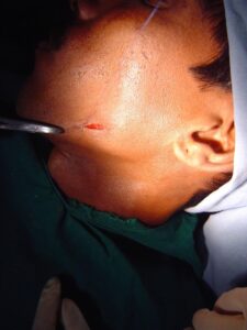
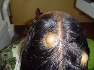
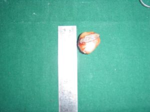
A soft tissue cyst is a fluid-filled sac that develops in the soft tissues of the oral cavity, such as the lips, cheeks, tongue, floor of the mouth, or gums.
These cysts are typically benign, slow-growing, and painless, but they can enlarge over time and interfere with oral functions.
Signs and Symptoms of Soft Tissue Cysts:
- Painless, dome-shaped swelling in the mouth
- Soft or firm lump under the mucosa
- Clear or bluish appearance (especially in mucoceles)
- Possible rupture leading to fluid drainage
- Discomfort or difficulty in speaking, chewing, or swallowing if the cyst enlarges.
Common Types of Soft Tissue Cysts:
- Mucocele: Forms due to trauma or blockage of salivary glands, commonly on the lips or floor of the mouth.
- Epidermoid/Dermoid Cyst: Develops from trapped epithelial cells, usually in the floor of the mouth.
- Lymphoepithelial Cyst: Appears as a small, yellowish lump in the soft tissues, often in the tongue or floor of the mouth.
Soft tissue cysts are usually diagnosed through clinical examination and imaging. In some cases, a biopsy may be needed to rule out other conditions. Treatment often involves surgical excision to prevent recurrence, especially if the cyst persists or causes discomfort.
Regular dental checkups help in early detection and management of soft tissue cysts, preventing complications and ensuring good oral health.
Ameloblastoma
Ameloblastoma is a rare, benign but locally aggressive odontogenic tumor that arises from the cells responsible for tooth development.
It most commonly affects the mandible (lower jaw), particularly the molar and ramus regions, but can also occur in the maxilla (upper jaw). Although it is slow-growing and painless, it can cause significant facial deformity and bone destruction if left untreated.
Signs and Symptoms of Ameloblastoma:
- Painless swelling or expansion of the jaw
- Facial asymmetry due to tumor growth
- Loosening or displacement of teeth
- Difficulty in chewing, swallowing, or speaking in advanced cases
- Occasionally, pain and infection if the tumor becomes secondarily infected.




Diagnosis and Treatment:
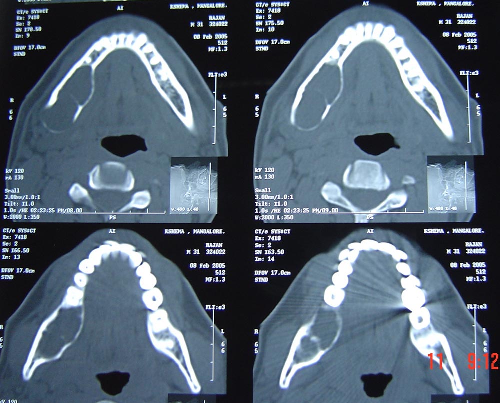
Ameloblastomas are typically detected through radiographic imaging (X-rays, CT scans, or MRIs), where they appear as soap bubble or honeycomb-like radiolucent lesions. A biopsy is necessary to confirm the diagnosis.
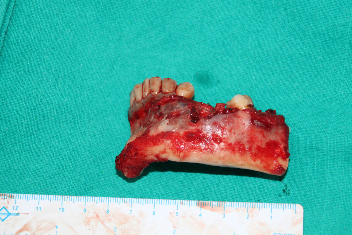
Treatment involves surgical removal, which may require resection of the affected jawbone to prevent recurrence. In some cases, reconstruction surgery may be needed to restore jaw function and aesthetics. Regular follow-ups are essential, as ameloblastomas have a high recurrence rate if not completely excised.
Early diagnosis and intervention are crucial to managing ameloblastomas effectively, preventing extensive bone loss and functional impairment.

Hemangioma
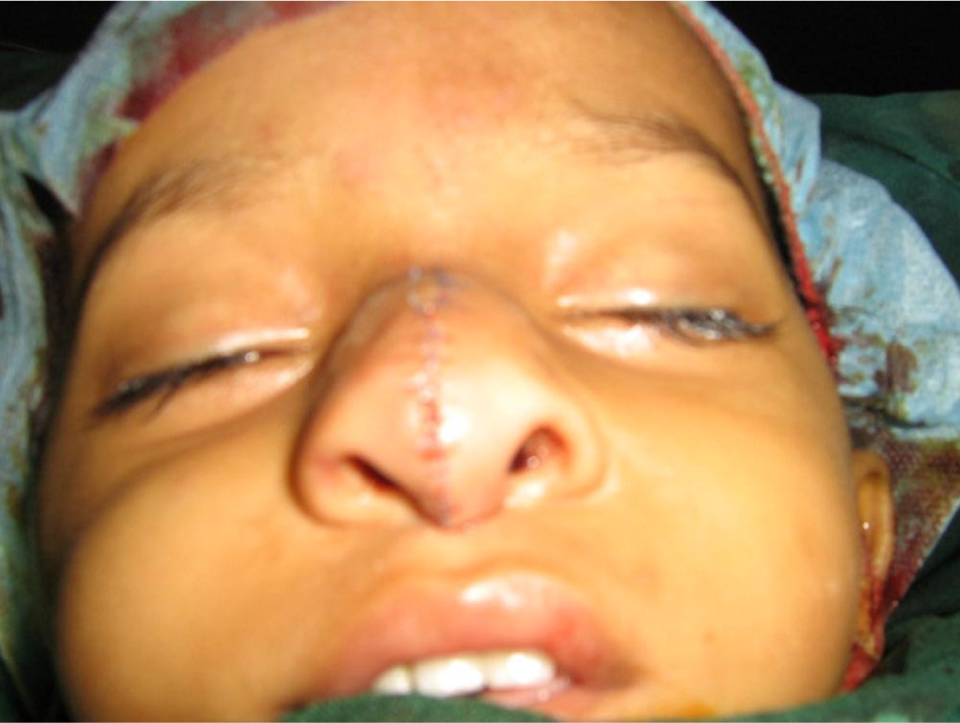
A hemangioma is a benign vascular tumor caused by an abnormal proliferation of blood vessels.
It can occur in the soft tissues of the oral cavity, including the lips, tongue, cheeks, and gums, or within the jawbone (intraosseous hemangioma). Hemangiomas are more common in infants and children but can also develop in adults.
Signs and Symptoms of Oral Hemangioma:
- Bluish-red or purplish lesion in the mouth
- Soft, compressible swelling that may increase in size over time
- Blanches (fades in color) when pressed due to blood flow changes
- Occasional bleeding, especially after trauma
- Pain or discomfort in rare cases when the lesion enlarges.
Diagnosis and Treatment:
Oral hemangiomas are diagnosed through clinical examination, Doppler ultrasound, and imaging techniques such as CT scans or MRIs to assess the extent of the vascular lesion.
Treatment depends on the size and location of the hemangioma. Small, asymptomatic hemangiomas may not require intervention and can be monitored. However, larger or problematic hemangiomas may need:
- Surgical excision (for accessible lesions)
- Laser therapy (to reduce size and bleeding risk)
- Sclerotherapy (injection of agents to shrink the lesion)
- Embolization (blocking blood flow to the tumor in complex cases)
Early diagnosis is essential to prevent complications, especially if the hemangioma interferes with oral functions or poses a risk of excessive bleeding. Regular monitoring and professional evaluation ensure proper management.
Dr. Gagan Sabharwal's clinic: Delhi
011-45033566
Fakeeh University Hospital, Dubai
+971-44144444
Jumeirah Clinic, Dubai
+1-80037569
